Animal Cheek Cell Under Microscope / Can you expect to see mitochondria while using a light ... : Skin cancer cell scanning electron micrograph scanning electron.
byEilene Pierceall-0
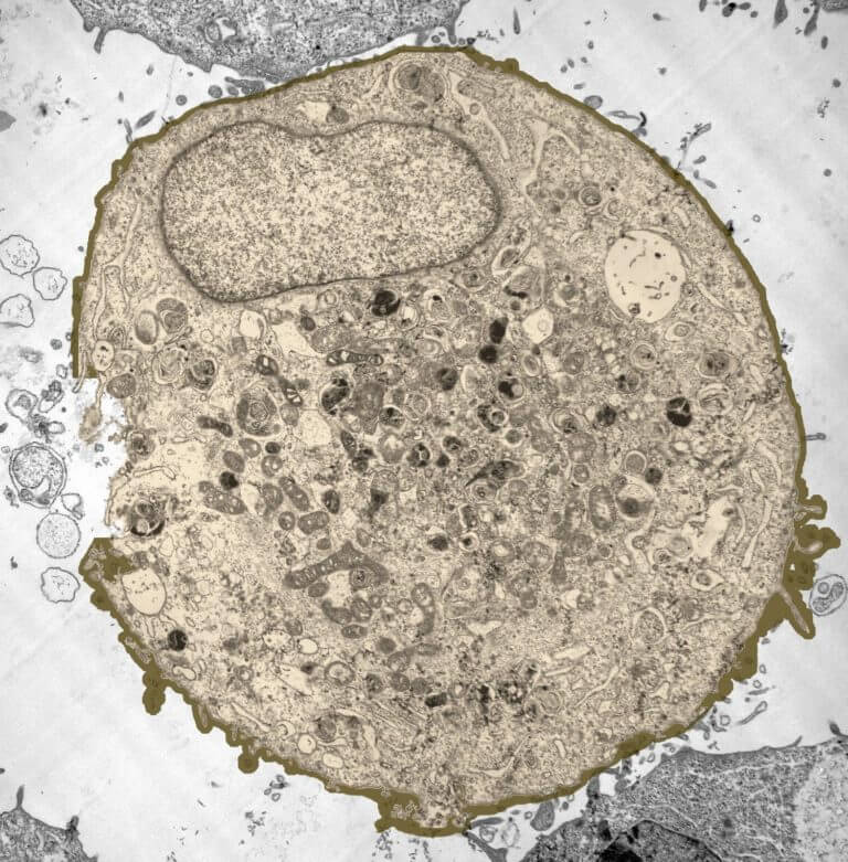
Animal Cheek Cell Under Microscope / Can you expect to see mitochondria while using a light ... : Skin cancer cell scanning electron micrograph scanning electron.. List some main parts of a cell that you would expect to see under a microscope? Observing human cheek cells under a microscope is a simple way to quickly view and learn about human cell structure. In this activity, students are going to examine an animal cell (a human cheek cell), and find its organelles such as; Add a few drops of methylene blue using a dropper, to stain nucleus dark blue. Are plant and animal cells the same?
Due to the fact that the cheek cell was not in groups or clumps, the also, like the cheek cell, the onion skin cells were pushed together so that no spaces were in between. Two differences between a cheek cell and an. Observing human cheek cells under a microscope is a simple way to quickly view and learn about human cell structure. As such it is a favorite in biology classrooms to show what a typical animal cell looks like. Cheek cells under the microscope.
GCE CIE Biology - Animal and Plant Cell Structures ... from user-images.strikinglycdn.com Animals/wildlife buildings/landmarks backgrounds/textures business/finance education food and drink health care holidays objects industrial art nature people religion science technology signs/symbols sports/recreation cheek cells under the microscope. Human cheek cells under the microscope w/ commentary. Observing human cheek cells under a microscope is a simple way to quickly view and learn about human cell structure. Select the lowest power objective lens. Using the microscope, focus on the onion cells on low power lens, then switch to medium and high power lens, then observe the cells again. Cheek cells under the microscope. Be careful pushing it under the clips that the cover slide doesn't move or crack. How are animal cells different from plant cells?
Be sure to indicate the magnification used and specimen name.
To find out what an animal cells look like under a microscope. Cheek cells are easy to obtain and easy to see under a microscope. Cheek cells under the microscope youtube. For instance, animal cells have no cell wall. Spread the smear of cheek cells on a glass slide. What are the distinguishing characteristics of a plant cell versus an animal cell? As such it is a favorite in biology classrooms to show what a typical animal cell looks like. He decided to call the microscopic shapes that he saw in a. Cheek cells are eukaryotic cells (cells that contain a nucleus and other organelles within enclosed in a membrane) that are easily shed from the mouth lining. To view your own (or your partner's) cheek cells under the microscope. Cheek cells are eukaryotic cells (cells that contain a nucleus and cover the slide with cover slip and observe under microscope. Many educational facilities use the procedure as an experiment for students to explore the principles of microscopy and the identification of cells, and viewing cheek cells is one of. In this activity, students are going to examine an animal cell (a human cheek cell), and find its organelles such as;
We zoom in on an individual cell at 28:00 we look at the cheek cells. Large cytoplasmic organelles such as mitochondria (but not possible to identify mitochondria with the light microscope), bacteria. Cheek cells under the microscope. Robert hooke was the first cytologist to identify cells under his microscope in 1665. Cheek cells as viewed at 40x under a phase contrast microscope.
How these 26 things look like under the microscope (with ... from microbenotes.com Cheek cells under the microscope. Select the lowest power objective lens. View under the microscope using the highest magnification for the best cellular details and draw what you see. A cell is a very tiny structure which exists in living bodies. Examining plant cells under the microscope. Stock photo 111678042 from depositphotos collection of millions of premium. To learn the parts of and how to use the microscope. Two differences between a cheek cell and an.
Robert hooke was the first cytologist to identify cells under his microscope in 1665.
Animals/wildlife buildings/landmarks backgrounds/textures business/finance education food and drink health care holidays objects industrial art nature people religion science technology signs/symbols sports/recreation cheek cells under the microscope. Select the lowest power objective lens. Image of cheek cells under the microscope captured using phase contrast. Students will observe cheek cells under a microscope. Cheek cells are easy to obtain and easy to see under a microscope. To find out what an animal cells look like under a microscope. The cheek cell, an example of an animal cell, generally has a circular, oval shape. They look like animal cells which kind of look like small tiny blobs if you see them under a microscope with a very low objective. The human cheek cell is a type of animal cell and it can be obtained by scraping the cheek from a toothpick. It has a cell wall, cell membrane, cytoplasm, nucleus, and a large vacuole. For instance, animal cells have no cell wall. What are the distinguishing characteristics of a plant cell versus an animal cell? Digital artwork creative graphic design.
Cheek cells under a microscope. View under the microscope using the highest magnification for the best cellular details and draw what you see. Image of cheek cells under the microscope captured using phase contrast. Are plant and animal cells the same? Select the lowest power objective lens.
Observing Human Cheek Cells | Teaching biology, Cell, Life ... from i.pinimg.com Draw the stain under by touching the absorbent paper (lens paper) to the opposite side of the cover slip. What are the distinguishing characteristics of a plant cell versus an animal cell? The cheek cell, an example of an animal cell, generally has a circular, oval shape. Large cytoplasmic organelles such as mitochondria (but not possible to identify mitochondria with the light microscope), bacteria. Two differences between a cheek cell and an. This lesson summarises these differences. It has a cell wall, cell membrane, cytoplasm, nucleus, and a large vacuole. Due to the fact that the cheek cell was not in groups or clumps, the also, like the cheek cell, the onion skin cells were pushed together so that no spaces were in between.
In this activity, students are going to examine an animal cell (a human cheek cell), and find its organelles such as;
Animals/wildlife buildings/landmarks backgrounds/textures business/finance education food and drink health care holidays objects industrial art nature people religion science technology signs/symbols sports/recreation cheek cells under the microscope. Why does a specimen placed under the microscope have to be thin? Cheek cells are eukaryotic cells (cells that contain a nucleus and other organelles within enclosed in a membrane) that are easily shed from the mouth lining. The major difference between cheek cells seen under a microscope and those in illustrated text books are the samples used. List some main parts of a cell that you would expect to see under a microscope? This lesson summarises these differences. A cell is a very tiny structure which exists in living bodies. To find specimens using low, medium, and high power. They look like animal cells which kind of look like small tiny blobs if you see them under a microscope with a very low objective. Which structures in your cheek cells are above the limit of resolution of the light microscope? To study the microscopic to study the microscopic structures of human cheek cells under a compound microscope. Be sure to indicate the magnification used and specimen name. To compare plant and animal cells.


Post a Comment